
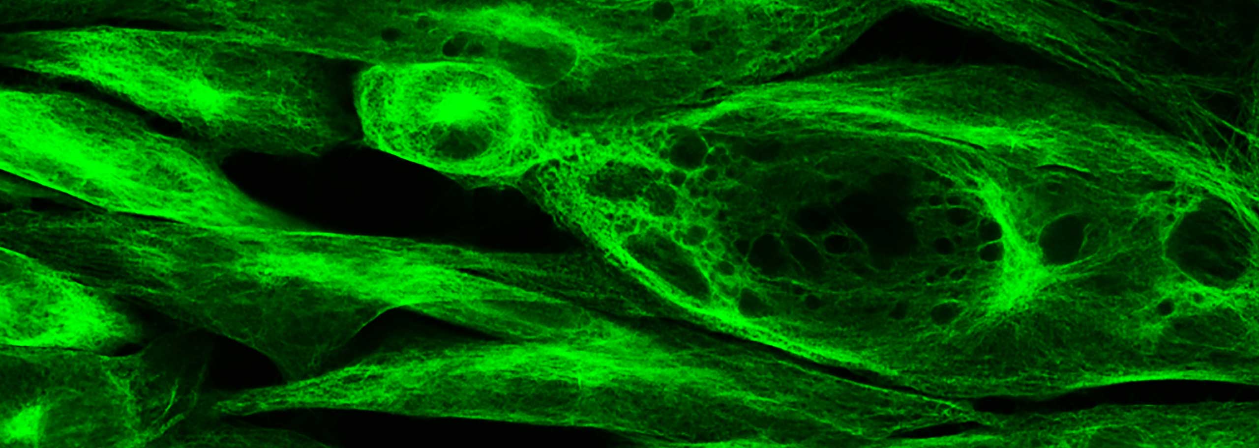
FRET Overview
FRET, or Forster Resonance Energy Transfer, is a phenomenon where closely matched pairs of fluorophores are used to determine spatial proximity in molecular and protein-protein interactions. Three criteria must be met for efficient FRET to occur between the donor and acceptor chromophores:
1. Strong spectral overlap between the emission spectrum of the donor and the excitation spectrum of the acceptor
2. Close proximity of the donor and acceptor chromophores (<10 nm)
3. Alignment of the transition dipoles of the two molecules
When these criteria are met, the energy is non-radiatively transferred from the donor to the acceptor. As a result, the donor's fluorescence intensity goes down (and it's excited-state lifetime is shortened) and the acceptor's fluorescence intensity goes up.
As described above, the donor and acceptor chromophores need to have a large spectral overlap, which, while creating the conditions for effective energy transfer, also results in spectral bleed through (SBT) and can be a problem in FRET measurements. Because excitation and emission spectra are very wide, there is bleed through in both directions. A small percentage of acceptor molecules can be directly excited by the donor's excitation, and simultaneously, a small percentage of donor emission can pass through the acceptor's emission filter.
The development of SBT correction techniques have been critical to the evolution of FRET as a useful and widely used application. These SBT correction techniques—which include software development, fluorescence lifetime imaging (FLIM) correlation, and photo bleaching techniques—have reached a degree of sophistication that improves the efficacy of FRET. Similarly, the development of microscopy techniques such as one-photon, two-photon (multiphoton), confocal, and TIRF are all contributing to the growing effectiveness and ease of FRET experiments.
Filter & Microscope Configurations for FRET
The filter components required for FRET experiments are not esoteric. As in any fluorescence microscopy application, an excitation filter is required for exciting the donor fluorophore, and a dichroic mirror is required for separating donor excitation energy from both donor and acceptor emission energy. Unlike typical fluorescence applications, however, two emission filters are required, one for the acceptor fluorophore, or FRET emission, and one for the donor fluorophore in order to correct for signal bleed thru. For the most part, the filter components used for FRET are identical to those used for epifluorescence applications, providing that the dichroic mirror transmits both emission wavelengths. More important than the selection of filters is an understanding of the physical configuration of the microscope hardware to be used in the FRET experiment. At issue are critical experimental variables, such as time and image registration. While the ideal setup may not be affordable or available to all researchers interested in FRET studies, it is nonetheless important to understand the pros and cons of the available hardware and filter set configurations.
1. Simultaneous Multi-view Microscope Configurations
For dynamic imaging of FRET due to protein-protein or other intermolecular interactions, the preferred setup allows the simultaneous visualization of both donor and acceptor signals. This arrangement is achieved with a series of dichroic mirrors and detectors arranged in a sequential manner such that different wavelengths are imaged onto each detector at the same time. The detectors are aligned so they are in perfect registration. Real-time corrections of bleed through can be accomplished using this method. Because the signals are acquired at the same time, it is the best method to use for imaging of live specimens. It is costly to set up, but robust because there are no moving parts in the system.
2. Emission Filter Wheels for Microscopes
The next best option is to use a filter wheel for fast-switching between donor and acceptor emission filters. When using a filter wheel in front of the detector, the filter cube is equipped with an excitation filter and dichroic mirror with the emission side left empty. When the donor and acceptor filters are loaded in the filter wheel, switching can be accomplished in ~50 ms which is fast enough for many biological processes. Because a single detector is used, this method is less expensive, but there may be subtle shifts in image registration due to movement of the filter wheel and variations between the filters.
3. Separate Filter Cubes
Without a multi-view attachment or emission filter wheel, researchers must fall back on a third hardware configuration for FRET, which involves using a separate filter cube for both donor and acceptor fluorescence. In this configuration, while each cube has an exciter, dichroic and emitter, it is extremely important to remember that the exciter and dichroic in both sets are identical. While this hardware configuration allows for the discreet collection of donor and acceptor fluorescence, it is slow (usually dependent on users explicitly changing filter cubes) and the most susceptible to subtle image registration shifts. It is most appropriate for fixed samples or those that are changing very slowly over time.